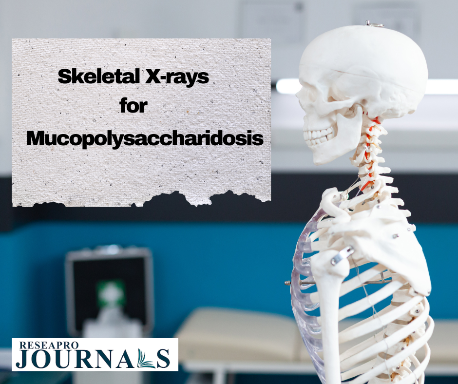
The clinical findings in children referred to the hospital for other ailments becoming evident, radiographs can offer hints for the diagnosis of MPS. As a result, children living in MPS-endemic areas may undergo X-ray screening to check for dysostosis multiplex.
What are the X-ray findings in mucopolysaccharidosis?
Paddle-shaped ribs, thick clavicles, wedge-shaped vertebral bodies with anterior breaking, platyspondyly, odontoid hypoplasia, lumbar gibbus, dorsal kyphosis, broad disc gaps, and spinal canal stenosis are among the distinctive radiographic findings.
What is dysostosis multiplex mucopolysaccharidosis?
We refer to the skeletal defects linked to MPSs as dysostosis multiplex (DM). These progressive alterations are most prominently observed in the severe manifestations of MPS VI (Maroteaux-Lamy syndrome) and MPS I (Hurler syndrome).
Common in MPS and ML, characteristic skeletal defects called dysostosis multiplex are caused by intra-lysosomal storage of glycosaminoglycans in cartilage, bone, and ligament cells. In MPS and ML, the hip joint is seriously impacted.
#mucopolysaccharidosis #MPS #X-ray #Radiographs